|
|
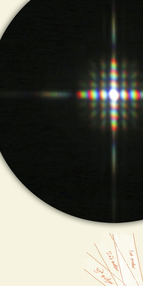
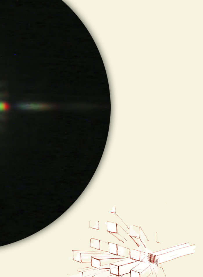
| Venus through a screen brightly Venus was low in the Calgary, Alberta evening sky when Alan Clark pictured it shining through the wire mesh screen of a newly installed window. The screen, a square mesh of wires 1.5mm apart has scattered Venus�s bright light into a colourful, grid-like diffraction pattern. This is a telephoto shot from a distance (500mm lens on a Canon 30D). Screen diffraction is not apparent close up unless the grid size is much smaller. ©Alan Clark, shown with permission |
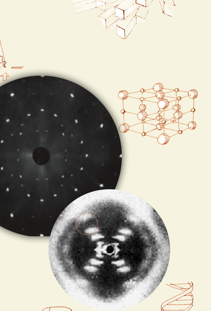
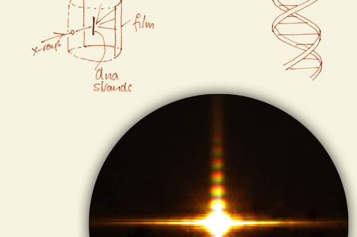
| The stuff of life At right: The epochal image made in 1951 by Rosalind Franklin and her student Raymond Gosling. She produced filaments of hydrated calf thymus DNA as would exist in a living cell. She reasoned that the molecules would be fairly well aligned in the thin strands and thus give decent x-ray diffraction images. The resulting pattern was the most informative ever obtained. From it she deduced that (1) DNA was helical (the cross shouts out �helix� and gives the helix angle). (2) The fine structure in the cross gave the helix pitch, (3) its diameter and (4) that there are 10 layer subunits to each helix turn. The missing diffraction spot (red circle) said (5) that there is a second co-axial helix present staggered 3/8 of a turn from the first. A double helix. All this from an unprepossessing blur! Key aspects of Rosalind Franklin's experiment can be done at home with an old ball-point pen and a laser pointer |
The window mesh is a two-dimensional diffraction grating. It sends out highly directed orders of diffracted light. The central, zero order, light is nominally undiffracted. Around it are successively higher orders, each one a a mini-spectrum. |
Salt is a face centered cubic array of positive sodium ions (small spheres) and negatively charged chloride ions. Convention shows them well separated but in reality they pack closely. |
|
|

| About - Submit | Optics Picture of the Day | Galleries | Previous | Next | Today |
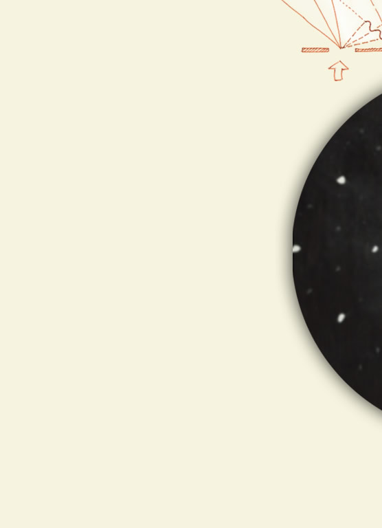
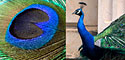
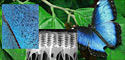
| 3-D gratings We see the action of 3-D gratings in the colours of a peacock�s tail and the vivid iridescent blues of a butterfly wing. On a much smaller scale, crystals act as 3-D gratings. Their immaculately ordered rows of atoms and ions scatter x-rays or neutrons (acting as waves) to form enigmatic spotty patterns that with some guile can be unwound to reveal the crystal�s structure. At right a Laue photograph of common salt (after Von Laue who first recorded x-ray diffraction by a crystal). A salt crystal is illuminated by a narrow x-ray beam of wavelength comparable to atomic dimensions. Each spot of the diffraction pattern is a diffracted beam of x-rays from planes of atoms of a particular direction. Analysis gives the atomic structure and the distances between the atoms/ions. |
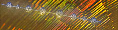    |
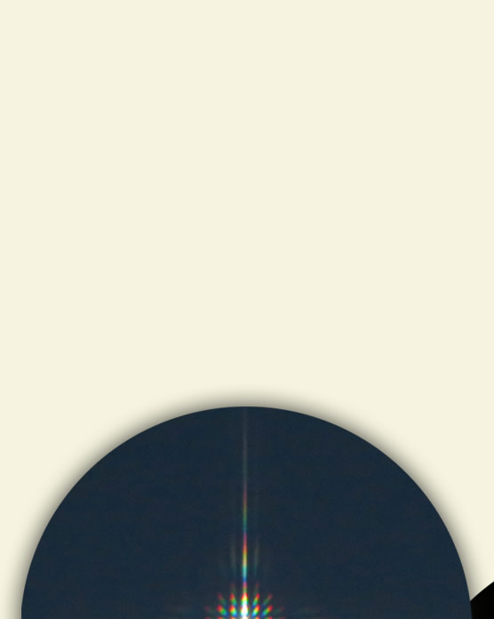
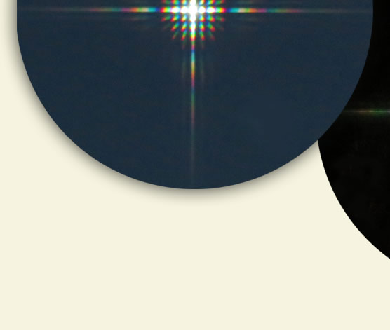
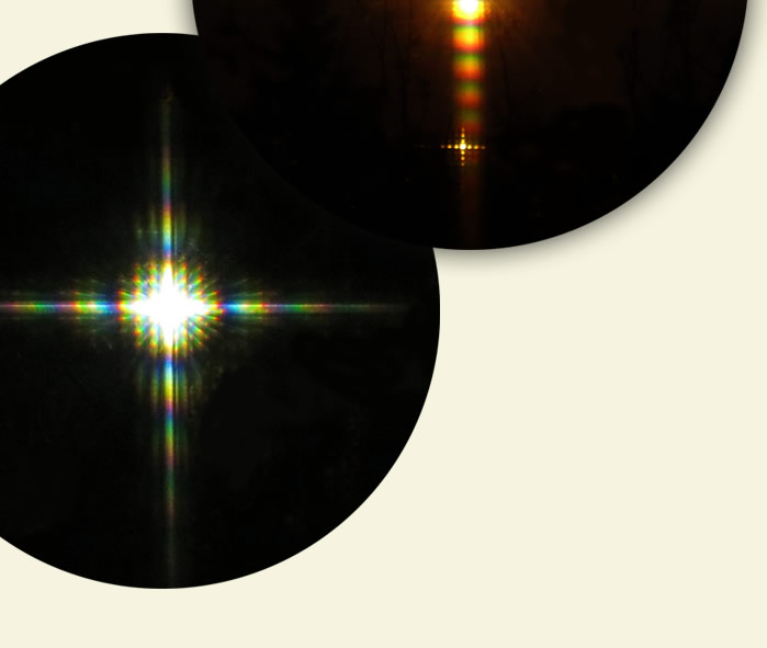
| Back to wire screens Left to right are Venus, a mercury street lamp and a high pressure sodium lamp. The wire mesh starts to reveal the discrete line spectrum of excited mercury vapour. |
Light is diffracted initially by a grating into all directions. Only some of it reaches any distance � the far field. The successful rays of the first order diffraction spectrum are in a direction that those from adjacent openings have a path difference of one complete wavelength. Second order spectrum rays come from rays having a path difference of two wavelengths from adjacent openings. And so on. Rays in other directions that have path lengths differing from a whole number of wavelengths eventually overlap and interfere destructively to give darkness. Only two openings are pictured. To have sufficient angular and thus spectral resolution a grating must have tens, hundreds or thousands of equally spaced openings. |